Home

Migration le golf client doppler normal Facture dachat Mettre quil sagisse

A Clinician's Guide to Tissue Doppler Imaging | Circulation

Doppler echocardiography - Wikipedia

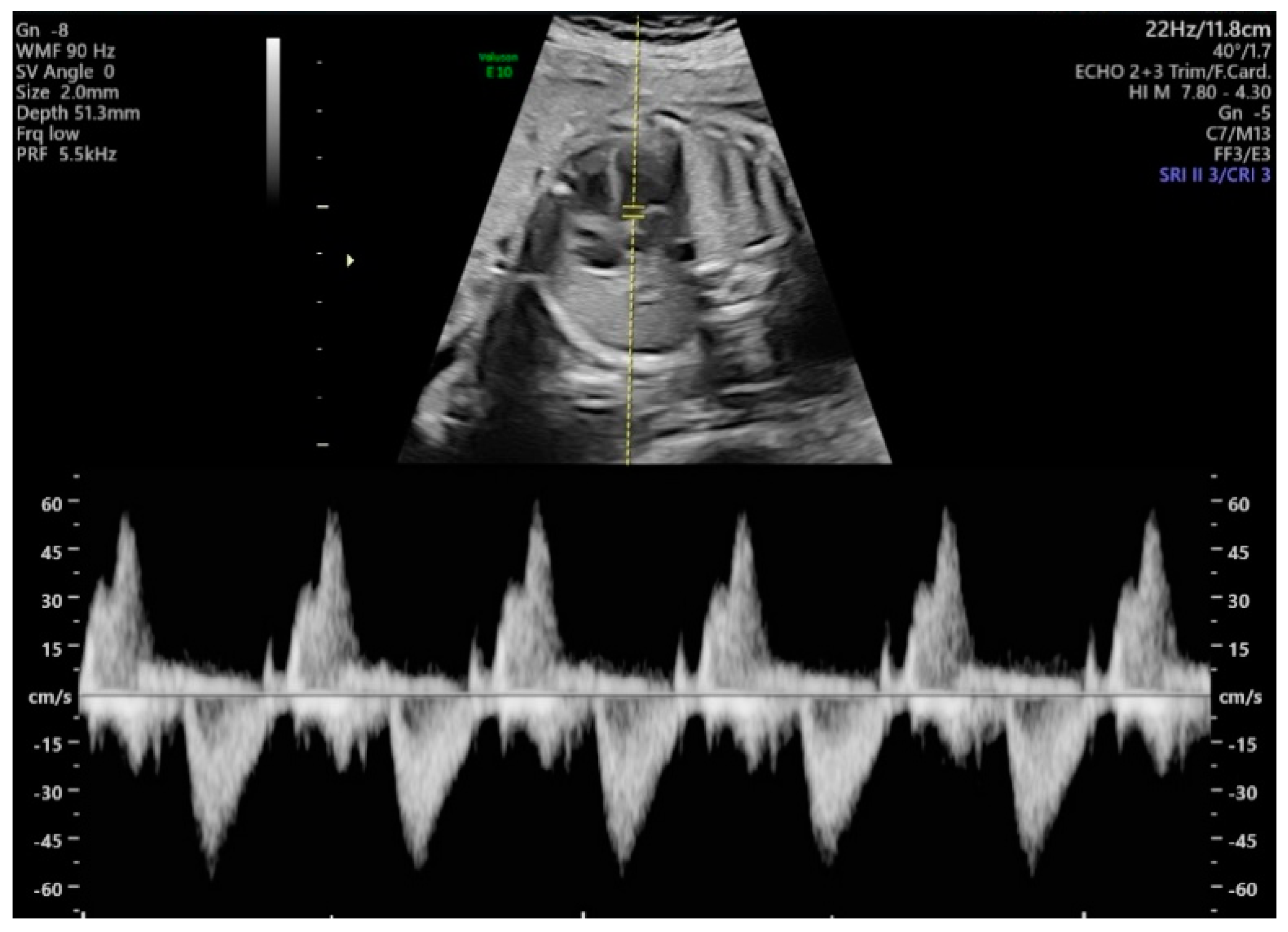
Diagnostics | Free Full-Text | Evaluation of the Fetal Left Ventricular  Myocardial Performance Index (MPI) by Using an Automated Measurement of  Doppler Signals in Normal Pregnancies
Diagnostics | Free Full-Text | Evaluation of the Fetal Left Ventricular Myocardial Performance Index (MPI) by Using an Automated Measurement of Doppler Signals in Normal Pregnancies
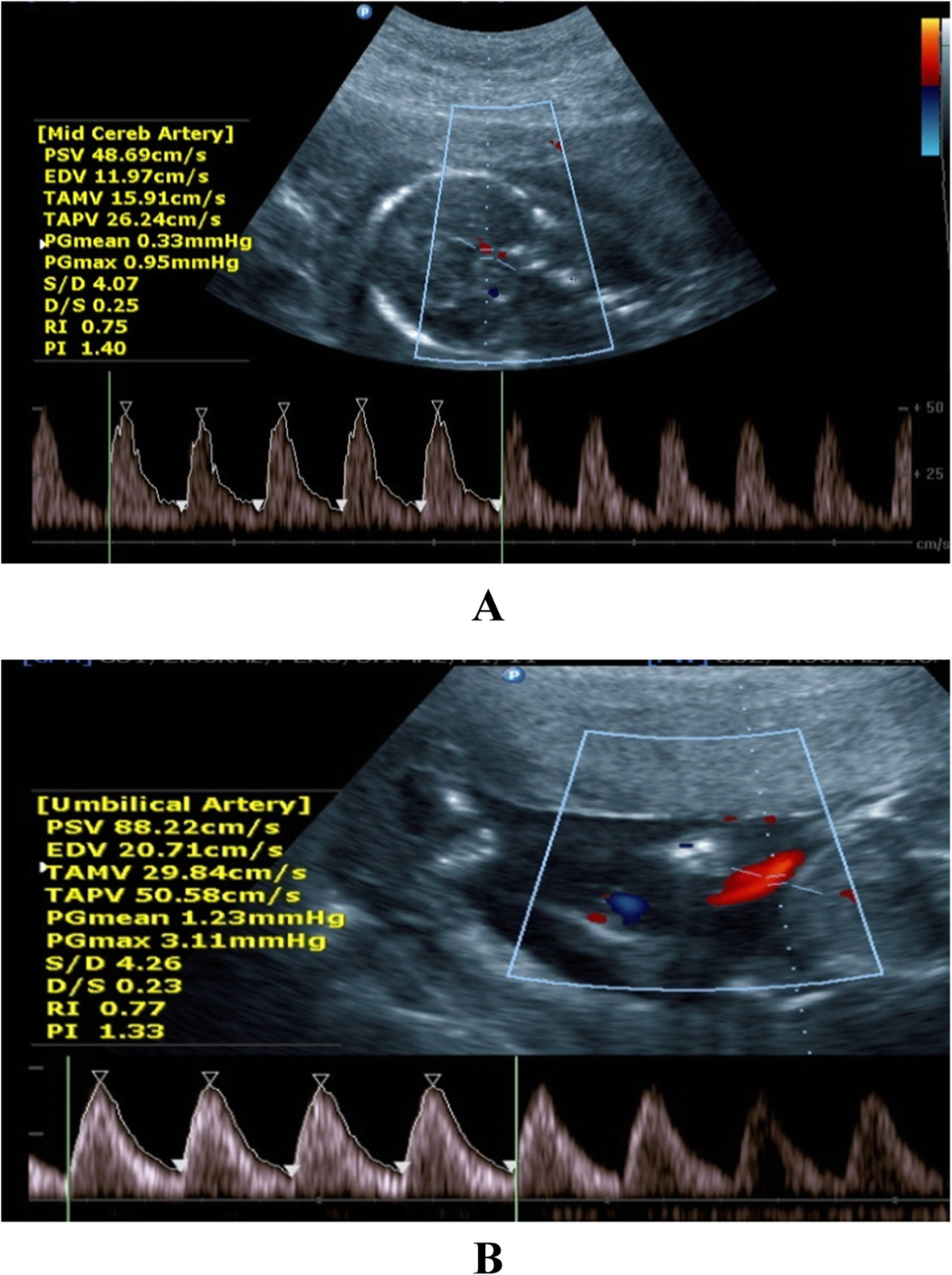
Ultrasound angiology reference standards of fetal cerebroplacental flow in  normal Egyptian gestation: statistical analysis of one thousand  observations | Egyptian Journal of Radiology and Nuclear Medicine | Full  Text
Ultrasound angiology reference standards of fetal cerebroplacental flow in normal Egyptian gestation: statistical analysis of one thousand observations | Egyptian Journal of Radiology and Nuclear Medicine | Full Text

UA Doppler ultrasound normal-600x400 - USF Health NewsUSF Health News

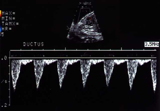
Fetal Ductus Venosus Doppler Ultrasound Normal Vs Abnormal Image  Appearances | Spectral Doppler USG - YouTube
Fetal Ductus Venosus Doppler Ultrasound Normal Vs Abnormal Image Appearances | Spectral Doppler USG - YouTube

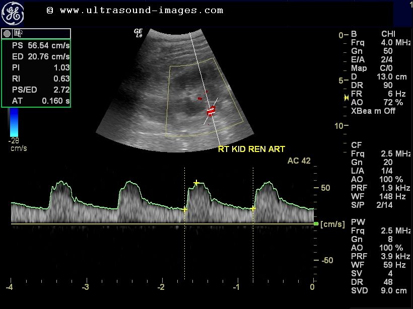
cochinblogs: Normal renal arterial color doppler study

A) Normal Doppler spectrum with spectral window (indicated by arrow).... |  Download Scientific Diagram
A) Normal Doppler spectrum with spectral window (indicated by arrow).... | Download Scientific Diagram
Liver Doppler Ultrasound - Pulsatility in Portal Vein Flow — @swissnephro

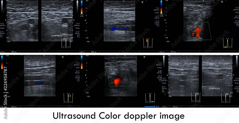
Ultrasound color doppler image:Normal doppler signal of superficial femoral  v. and popliteal v. bilaterally.No sign of thrombus in the veins. Stock  Illustration | Adobe Stock
Ultrasound color doppler image:Normal doppler signal of superficial femoral v. and popliteal v. bilaterally.No sign of thrombus in the veins. Stock Illustration | Adobe Stock

Doppler ultrasound assessment of the ductus venosus in the normal fetus  between 20 and 40 weeks gestation in the Pakistani population - Syed Amir  Gilani, Amber Javaid, Alsafi Abdella Bala, 2010
Doppler ultrasound assessment of the ductus venosus in the normal fetus between 20 and 40 weeks gestation in the Pakistani population - Syed Amir Gilani, Amber Javaid, Alsafi Abdella Bala, 2010

Umbilical Artery Doppler Ultrasound Normal Vs Abnormal Image Appearances |  Spectral Doppler USG - YouTube
Umbilical Artery Doppler Ultrasound Normal Vs Abnormal Image Appearances | Spectral Doppler USG - YouTube

Normal Doppler Spectral Waveforms of Major Pediatric Vessels: Specific  Patterns | RadioGraphics
Normal Doppler Spectral Waveforms of Major Pediatric Vessels: Specific Patterns | RadioGraphics

A Radiologist's Guide to the Performance and Interpretation of Obstetric  Doppler US | RadioGraphics
A Radiologist's Guide to the Performance and Interpretation of Obstetric Doppler US | RadioGraphics

Umbilical arterial Doppler assessment | Radiology Reference Article |  Radiopaedia.org
Umbilical arterial Doppler assessment | Radiology Reference Article | Radiopaedia.org

Umbilical Artery Doppler – Sonographic Tendencies

Fetal Echocardiography Homepage

Liver Transplant Ultrasound | IAME

Doppler ultrasound assessment of the ductus venosus in the normal fetus  between 20 and 40 weeks gestation in the Pakistani population - Syed Amir  Gilani, Amber Javaid, Alsafi Abdella Bala, 2010
Doppler ultrasound assessment of the ductus venosus in the normal fetus between 20 and 40 weeks gestation in the Pakistani population - Syed Amir Gilani, Amber Javaid, Alsafi Abdella Bala, 2010